Description
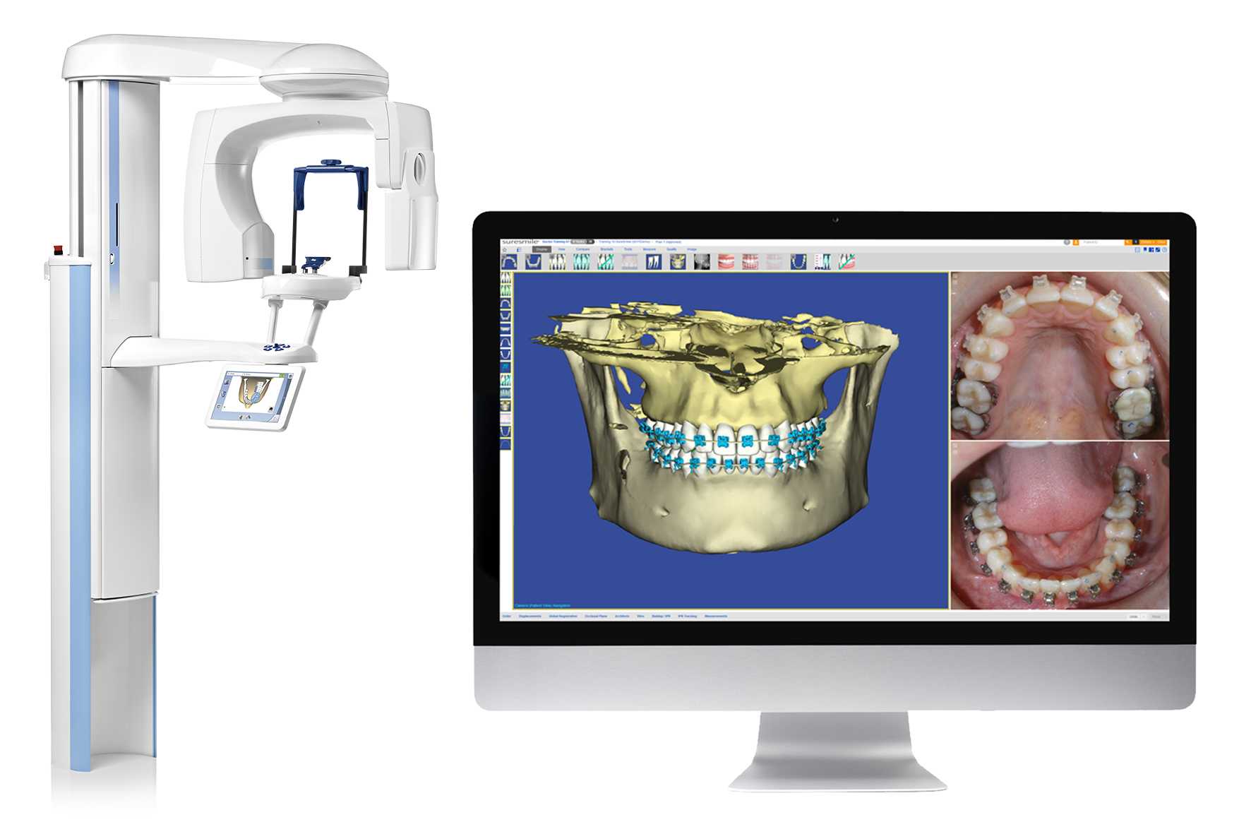
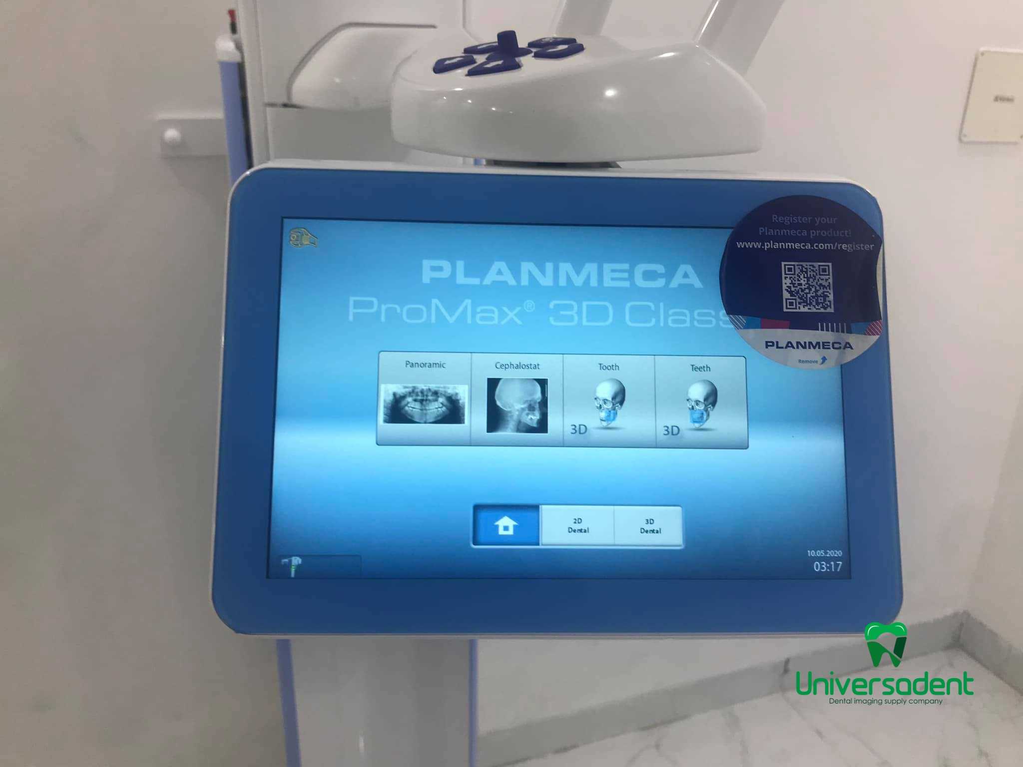
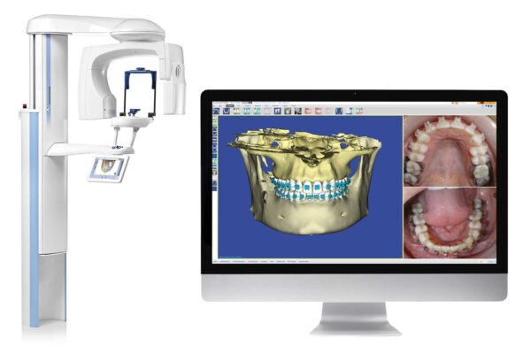
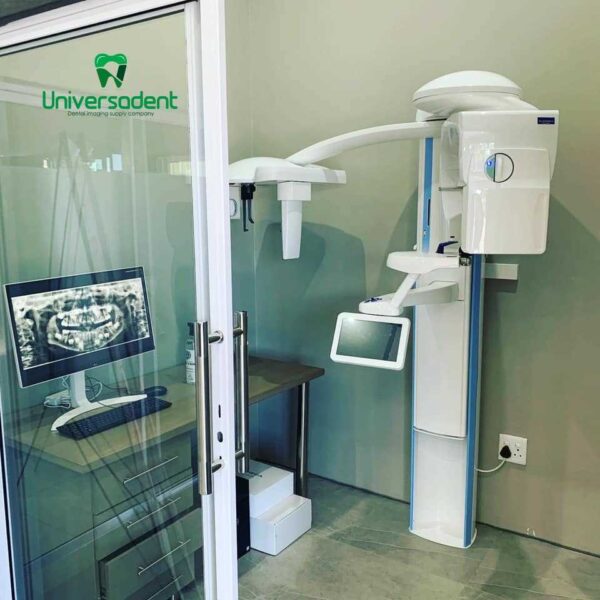
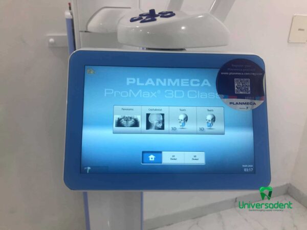
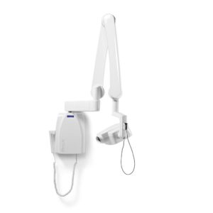
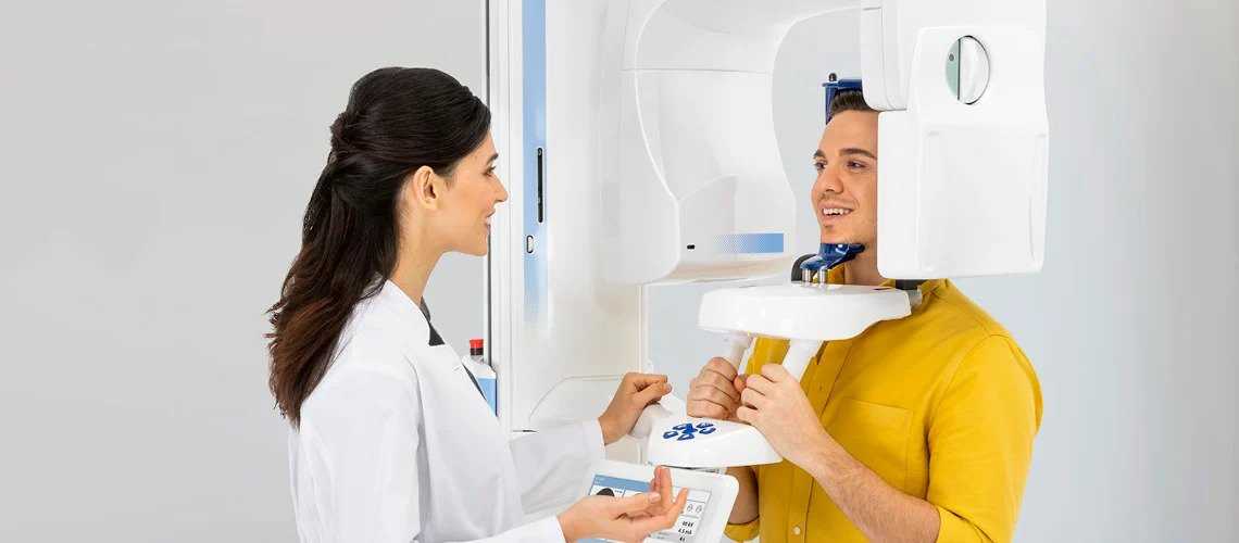
Planmeca ProMax 3D Classic unit is designed to obtain complete information on your patient’s anatomy in the minutest detail. This intelligent and multipurpose X-ray unit provides digital panoramic, cephalometric and 3D imaging as well as 3D photos and 3D model scans.
The Planmeca ProMax 3D Classic CBCT is one member of the ProMax family. It provides coverage of the entire dentition, making it an excellent option for full arch dental 3D imaging needs.
Planmeca ProMax 3D is an all-in-one device series. Three different types of 3D imaging, as well as panoramic, cephalometric and external bite imaging. These multifunctional units can meet all your maxillofacial imaging needs.
Planmeca ProMax 3D X-rays allow imaging in the unique Planmeca Ultra Low Dose protocol mode, which allows CBCT acquisition with even lower patient radiation exposure than standard 2D panoramic imaging. This groundbreaking tomography protocol is based on intelligent 3D algorithms developed by Planmeca and provides detailed patient anatomical data at a very low dose.
Planmeca CALM
Patient movement can cause problems when taking 3D images. Planmeca’s CALM algorithm allows for the removal of artifacts caused by movement- leading to a much sharper final image without having to re-take images.
Low Dose Images
This is a standard feature on all Planmeca’s 3D units. Planmeca’s Ultra Low Dose imaging protocol allows you to capture CBCT images with an even lower dose than panoramic images.
All-in-one Imaging Software
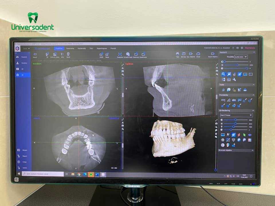
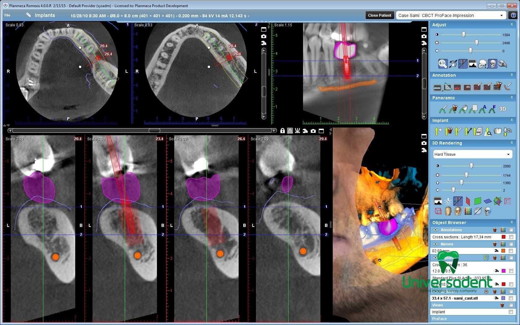
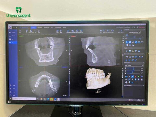
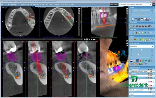
The Planmeca Romexis imaging software supports all 2D and 3D imaging as well as CAD/CAM work. The software offers an extended range of tools for dental clinics of all sizes.
Advantages of 3D model scanning
Digital models save space
3D digital models are stored in the Planmeca Romexis® database in standard STL format, which reduces the need to make or maintain physical plaster casts.
Create your virtual patient
The scanned 3D model can be superimposed on CBCT data, creating a virtual patient and helping you with all your clinical and treatment planning needs. The combined data set provides an artifact-free model of your patient’s dentition including bone, crowns, and soft tissue. This offers valuable new options for implant planning, surgical guide manufacturing, orthodontic purposes, and orthognathic surgery.
Endodontic imaging mode for Planmeca ProMax® 3D family
We are proud to introduce a new imaging mode that is specially designed for endodontic studies. The new imaging mode is available for all new Planmeca ProMax® 3D family units and provides perfect visualization of even the finest anatomical details.
Thanks to our intelligent Planmeca AINO™ noise removal and Planmeca ARA™ artifact removal algorithms, the result is noise-free and crystal-clear images. The new imaging mode is ideal for endodontics and other cases with small anatomical details, such as ears.
Stand out with color
Complement the splendid design of your Planmeca ProMax® 3D X-ray unit by giving it a personal touch with your favourite colors. Select the perfectly matching shades from our exquisite and inspiring collection and create the looks of your dreams!
Benefits:
Hi-tech:
- Perfect resolution and patient dose level always follows the ALARA principle (as little as possible)
- Optimal volume size and location for each clinical trial
- Dedicated imaging protocols for dental and ENT applications
Easy use:
- Convenient patient positioning and unsurpassed comfort
- True all-in-one X-ray machines for not only 3D imaging but also 2D panoramic and cephalometric imaging
- Ease of use
- Planmeca Romexis® software
- Mac OS and Windows compatible
Key Features:
Extended Volume Size
Planmeca ProMax 3D Classic offers an exclusive extended volume diameter size. It is ideal for capturing the full dentition with one scan and increases the maximum diameter from Ø80×80 to up to Ø110×80 mm.
See Even the Finest Details
Planmeca’s endodontic mode offers an extremely high resolution with a voxel size of 75 μm. Optimal for capturing even the smallest details, the mode enables precise diagnostics and treatment planning.
Planmeca CALM
Planmeca’s CALM movement correction algorithm helps to remove movement artifacts from CBCT images meaning you don’t have to re-take images which takes up time, energy, and money.
Ideal for Imaging Braces
Planmeca ProMax 3D Classic’s Braces imaging protocol is ideal for orthodontics, as images captured using it show metal brackets with true precision. The unit has been certified for use with the SureSmile treatment system.
Technical characteristics of the Planmeca ProMax 3D Classic tomograph:
- X-ray tube Toshiba D-054SB
- Focal spot size 0.5 x 0.5 mm, per IEC 6036
- General filtration min. 2.5 mm Al + 0.5 mm Cu
- Anode voltage:
- panoramic mode 54 – 84 kV
- 3D mode 50-90 kV
- Anode current:
- panoramic: 1 – 16 mA
- three-dimensional: 1-14 mA
- Irradiation time 2.8 – 12 sec,
- Scan time 14 – 35 sec
- Magnification 1.57
- Single rotation 200 °
- Overall dimensions (WxDxH): 97 × 125 × 156–2385 cm.
- Chin level 96-178 cm
- Weight (net) 113 kg.
- SmartPan ™ system
- Implant planning module
- FOV Ø110 x 80 mm
- 2 Planmeca Romexis® 3D Advanced licenses (one basic + one additional in the implant planning module)
Digital 3D sensor specifications:
- Amorphous silicon flat panel with Cls scintillator
- Pixel size 127 μm
- Active surface 13×13 cm
- Sensor resolution 5 line pairs / mm
- Ethernet interface.
Include :
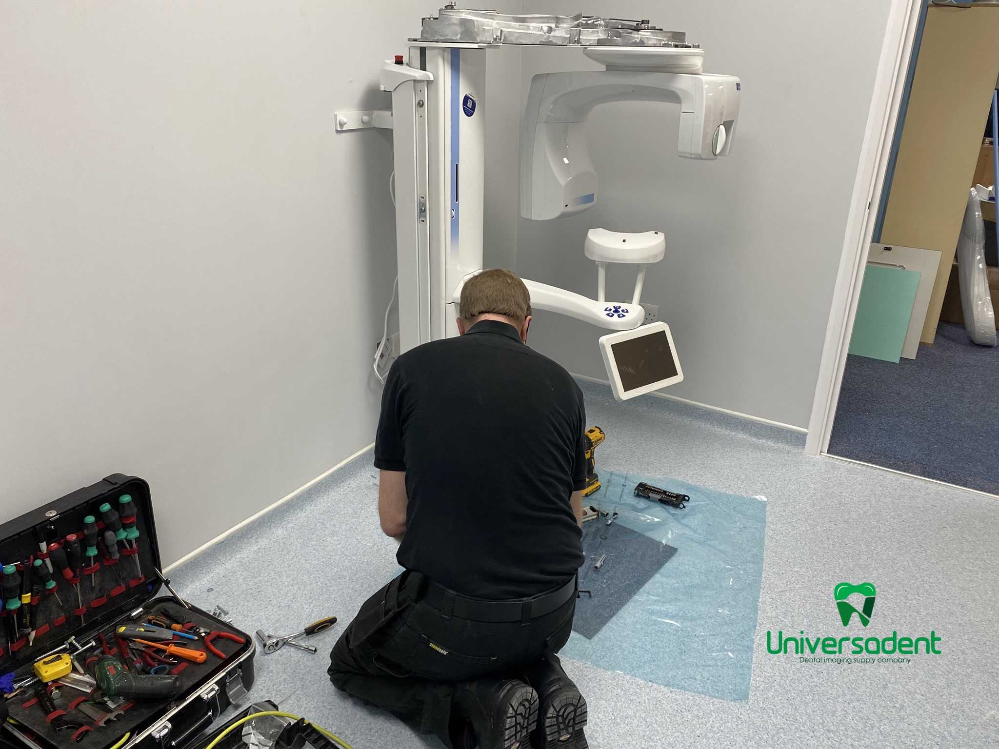
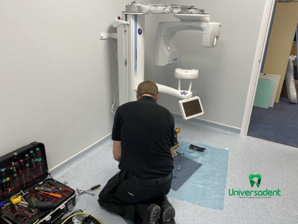
- Installation
- a comprehensive onsite service and replacement parts warranty
- unlimited support from the Help Desk.
- CEPH
- CONE BEAM
- Training
- Warranty
Reference Library
Planmeca Promax 3D Classic Manufacturer Information
Planmeca Promax 3D Classic Manufacturer Information Brochure

